About our instruments
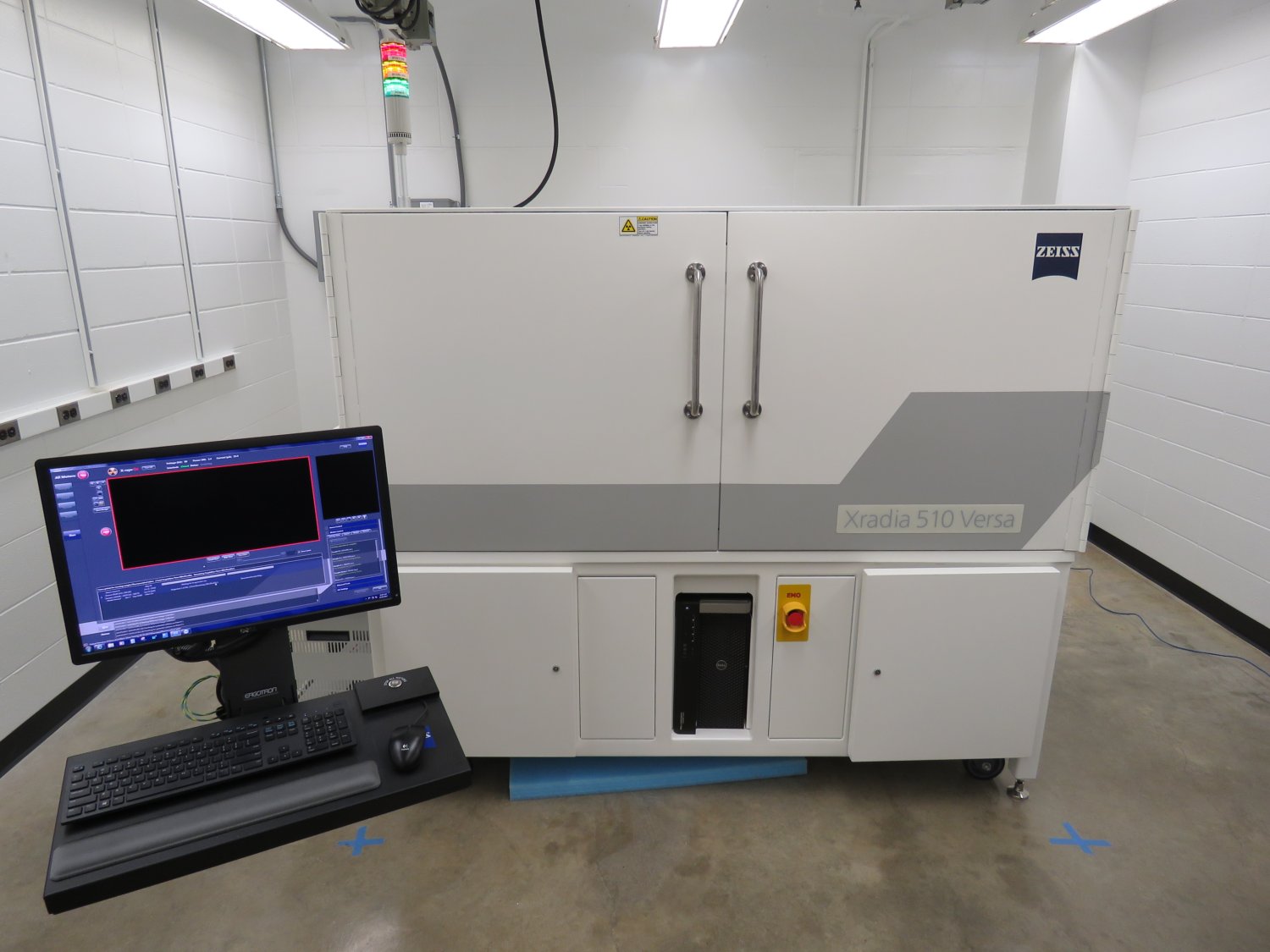
The Zeiss Xradia 510 Versa 3D X-Ray Microscope and Workstation located in the Department of Physics and Astronomy, room G48
The Zeiss Xradia 510 Versa is a versatile 3D X-Ray Microscope that provides excellent flexibility for 3D submicron imaging by allowing a wide range of sample sizes while maintaining high resolution even at large working distances providing users with high-quality 3D image data. The Scout + Scan and Zoom feature extend the non-destructive imaging capabilities by allowing users to zoom in to the exact location of features of interest. The system is built to support in-situ deformation, acoustic emission monitoring, and fluid** experiments and provides an excellent investigation tool for dynamic processes through 2D X-Ray imaging.
At the Purdue University 3D XRM Facility we also customize sample holders to ensure we can achieve the best scan parameters and proper scan positioning which is important when imaging dynamic processes and/ samples before and after some process (i.e. deformation, fluid movement, damage to microstructure, etc.)
Zeiss Xradia 510 Versa Technical Specifications |
|
|---|---|
| X-Ray Source (Voltage Levels) | 30 to 160 kV (10W maximum output) |
| Spatial Resolution | 0.7 μm |
| Minimum achievable Voxel (at max. magnification) | 70 nm |
| Detector Objectives (Magnification Lenses) | 0.4X, 4X, 20X |
| Source filters | High and Low Energy |
| Manual filters | |
| Sample Stage (motion) | Rotation around Y-axis (-180° to +180°) |
| Stage Load capacity | 15 kg |
| Sample stage Travel (in X, Y, Z) | 50, 100, 50 mm |
| Source travel (Z-axis) | 212 mm |
| Detector Travel (Z-axis) | 334 mm |
| Zeiss recommended sample size limit | 300 mm (Interior Scans) |
| In-situ system temperature | ~25-28°C |
| Scanning Modes | Normal, Wide Field, Vertical Stitching, WFST |
| Phase contrast can be optimized for low Z materials | |
| The systems Z-axis is defined to be along the X-Ray beam path | |
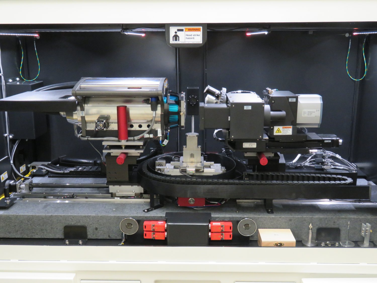
Inside the Zeiss Xradia 510 Versa 3D X-Ray Microscope showing the source (left) & receiver (right)
Vertical Stitching, Wide Field Mode (WFM) & Wide Field Mode (WF-St) Stitching
The 3D X-Ray microscope offers additional variability through the vertical stitching, wide field mode, and wide field mode stitching features. With these capabilities, large samples with an extended length field of view or even a standard field of view can be imaged in segments with higher resolution in single tomographies and then stitched together. With this technique, we gain a larger laterally or vertically extended field of view: approximately twice as wide as in the standard mode and three times larger for a 3D volume (for a wide field of view) and for vertical stitch the segments can be any number with a data set of a corresponding size. Combining both the WFM with the existing Vertical Stitching feature, Zeiss offers researchers the opportunity to image large samples that are both wider and taller than the standard field of view usually allows.
Scout - Scan & Zoom
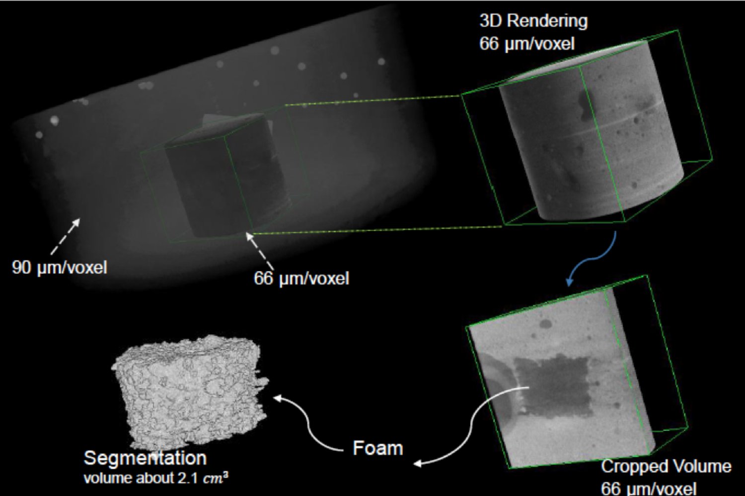
Example of Scan & Zoom Capabilities of the 3D X-Ray Microscope and segmentation (bottom left) on a 10-inch cement core
The Scan and Zoom feature provides a great opportunity for users who require high-resolution 3D imaging data but only at a certain position in a particular sample. In such cases, we recommend the Scan & Zoom option. Which involves first performing a large FOV of view scan at a good resolution, then locating the feature and/ area of interest and using these coordinates to set up a high-resolution scan. Once the scans are complete, the high-resolution image data can now be registered to the large FOV dataset thereby allowing users to show the exact location out of the large FOV data that was imaged and illuminate features that were not visible in the lower resolution data.
Image Analysis Workstation
Available Software for Image Analysis
- ORS DragonFLY Pro.
-
DATA IMPORT Dragonfly is the ideal framework for integrating data from multiple sources into a single environment, whether your images come from leading microscope and imaging hardware vendors or RAW files produced by academic software. Image stacks (.tif, .tiff, .jpeg, .jpg, .png, .bmp, .dib extensions). DICOM image files (.dcm extension). MRC (.mrc extension), REK (.rek extension), TXM (.txm extension), CZI (.czi extension). RAW data (.raw and .pic extensions) with or without header files (.dat extension). Analyze 7.5 files (.hdr extension with accompanying .img extension). VISUALIZATION Dragonfly provides a powerful set of image review and interactive inspection tools that let you gain detailed insight into your 3D data. ANNOTATIONS AND MEASUREMENTS Tools to add annotations to 2D and 3D views and to extract measurements from image data and processed objects include the following: IMAGE PROCESSING (GEOMETRY) Post-processing functions available in Dragonfly include data restructuring, registration, and 2D, 3D, and 4D stitching. IMAGE PROCESSING (FILTERING) Dragonfly's extensive image processing toolkit features filters to smooth images, sharpen edges, correct uneven shading, extract features, and more. With selectable 2D and 3D settings and user-specified kernel sizes, you can combine multiple filters together as a workflow, which can be saved and applied to a batch of images for automated processing. SEGMENTATION Image segmentation and intuitive masking operations in Dragonfly let you quickly identify and label regions of interest to gain detailed insights into material structures and properties. QUANTIFICATION Whether analyzing pores, fibers, grains, phases, or anything else, Dragonfly's quantification and analysis tools give you powerful options for counting, measuring, and characterizing image features. 3D MODELING Create meaningful and informative visualizations of polygonal models, vector fields, and spatial graphs generated from image data and segmented objects, or from simulations. PRESENTATION AND ORGANIZATION Reveal key insights and share your research with annotated high-resolution screenshots and tell dynamic stories with easy-to-produce animated sequences that can be exported as HD video files or uploaded to the web. EXTENSION AND AUTOMATION Dragonfly’s 3D analysis workflows can be customized with Python or extended with custom add-ons that implement specific tasks and workflows to suit your research objectives. See ORS DragonFLY Features page for more information
*Academic Users can obtain an educational copy of ORS DragonFLY. However much of the features of Pro will not be available and you will not be able to upload .txrm files. It is a good practice to always save the tiff stack of your data.
-
- FIJI - Extremely useful commonly used open-source image processing software