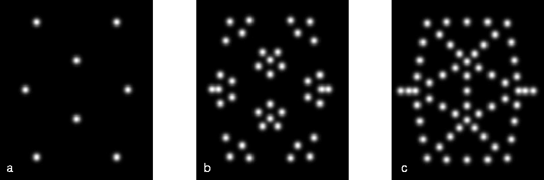
A few simulated FIM micrographs from the icosahedral cluster are shown in Fig. 6. In this series of simulated field-ion micrographs, the shell thickness t is systematically increased to allow successively more of the surface atoms on the icosahedral cluster to become evident. This calculation mimics the effects of increasing the electric field at the surface of the cluster. By referring to Fig. 4, the one-to-one correspondence between the atomic positions on the cluster surface and the simulated field-ion images become clear. Atoms missing from the cluster or the presence of additional atoms in the guise of partially filled shells should become evident in subsequent field-ion experiments.
Figure 6: Comparison of simulated FIM images from a 309-atom
icosahedral cluster (see Fig. 4) for varying shell thicknesses.
a) t = 0.75Å, b) t = 1.00Å, c) t = 1.50Å.
A simulated image of the truncated-octahedra cluster (see Fig. 5) is shown in Fig. 7. In contrast to the icosahedral cluster, a majority of the atoms on the surface of the truncated octahedral cluster are visible in the FIM micrograph. As is evident from these simulations, it is a simple matter to distinguish between an icosahedral cluster and a truncated octahedral cluster on the basis of their field-ion images. This ability to unambiguously determine atomic structure from the micrographs makes FIM an excellent choice for studying cluster structure and orientation.

Figure 7: Simulated FIM micrograph of the 201 atom truncated-octahedral
cluster shown in Fig. 5.
An attempt to compare a simulated field-ion image to the experimental image shown in Fig. 2 is worthwhile. A computer program was written to allow a rotation of the clusters through any combination of standard Euler angles.[20] By rotating the icosahedral cluster in this way and studying the resulting field-ion image, it is possible to find many orientations which match the quasi five-fold symmetry evident in Fig. 2. An example is plotted in Fig. 8. A detailed comparison between this simulation and the experimental FIM image is discussed in more detail elsewhere.[21]
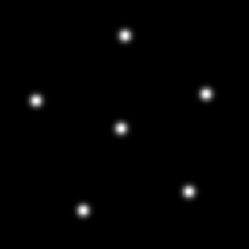
Figure 8: Simulated FIM image from a 309 atom icosahedral cluster
viewed from an orientation
approximating the experimental image shown in Fig. 2.