The principles governing the field-ion microscope have been known for
quite some time.[9, 10] Briefly, the FIM consists of
an electrochemically etched [11] metallic tip with an end
radius of about ![]() 50 nm, which is placed inside a vacuum chamber and
aimed at a fluorescent screen or multi-channel plate (MCP) required for
imaging. A strong electric field of order
50 nm, which is placed inside a vacuum chamber and
aimed at a fluorescent screen or multi-channel plate (MCP) required for
imaging. A strong electric field of order ![]() V/m is
achieved by applying about 10 kV between the tip and screen. Inert gas
(typically H
V/m is
achieved by applying about 10 kV between the tip and screen. Inert gas
(typically H ![]() or He) admitted to the chamber is then preferentially
ionized near the tip by the high electric field. Since some surface
atoms on the tip protrude slightly more than nearby adjacent atoms, a
local enhancement of the electric field results near these protruding
atoms. As a result, gas atoms are preferentially ionized at these
sites. After ionization, the gas atoms are accelerated towards the
screen by the applied potential difference, producing an image of the
surface atoms on the tip.
or He) admitted to the chamber is then preferentially
ionized near the tip by the high electric field. Since some surface
atoms on the tip protrude slightly more than nearby adjacent atoms, a
local enhancement of the electric field results near these protruding
atoms. As a result, gas atoms are preferentially ionized at these
sites. After ionization, the gas atoms are accelerated towards the
screen by the applied potential difference, producing an image of the
surface atoms on the tip.
The field-ion technique can provide interesting structural information about nanometer-size clusters if a cluster can be captured on the apex of a conventional field-ion tip. We have already demonstrated that it is possible to deposit clusters on a tip and to image the position of the surface atoms of the supported cluster using field-ion techniques. [4, 6, 7] A schematic of the apparatus required is shown in Fig. 1. One way to implement this experiment is to attach a UHV, differentially pumped vacuum chamber housing the tip to a cluster beam port. Provisions to allow a precision alignment of the tip with respect to the cluster beam must be included.
The deposition of clusters onto the apex of field-ion tips has met with
considerable success by simply placing the tip in a cluster beam. It
is easy to estimate that for a tip with radius of ![]() 50 nm, a cluster beam
flux of
50 nm, a cluster beam
flux of ![]() is required to
capture one cluster in the vicinity of the tip in one minute. The
deposition of a cluster is signaled by the appearance of a bright spot
in the field emission pattern as viewed on the fluorescent screen,
indicating the presence of an object with a small radius of curvature
on the tip.
is required to
capture one cluster in the vicinity of the tip in one minute. The
deposition of a cluster is signaled by the appearance of a bright spot
in the field emission pattern as viewed on the fluorescent screen,
indicating the presence of an object with a small radius of curvature
on the tip.
When this event is observed, the cluster beam can be switched off. The
tip must then be cooled to cryogenic temperatures in order to take full
advantage of the high resolution of the FIM. An example of a FIM
micrograph from a ![]() 2 nm Au cluster is shown in Fig.
2.[4] Each bright spot represents the position
of an atom on the surface of the cluster. The size of the spots are
related to the details of the experiment and the exposure time used to
photographically record the field-ion image. The quasi five-fold
symmetry of the field-ion pattern reflects the underlying symmetry of
the cluster while the relative positions of the spots with respect to each
other contains valuable information about the position of atoms on the
surface of the cluster.
2 nm Au cluster is shown in Fig.
2.[4] Each bright spot represents the position
of an atom on the surface of the cluster. The size of the spots are
related to the details of the experiment and the exposure time used to
photographically record the field-ion image. The quasi five-fold
symmetry of the field-ion pattern reflects the underlying symmetry of
the cluster while the relative positions of the spots with respect to each
other contains valuable information about the position of atoms on the
surface of the cluster.
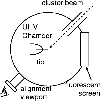
Figure 1: Generic FIM experiment for studying supported nanoscale clusters. A potential
difference between the tip and screen promotes electron emission from any cluster with small
radius of curvature and signals the deposition of a cluster on the apex of the field emission tip.
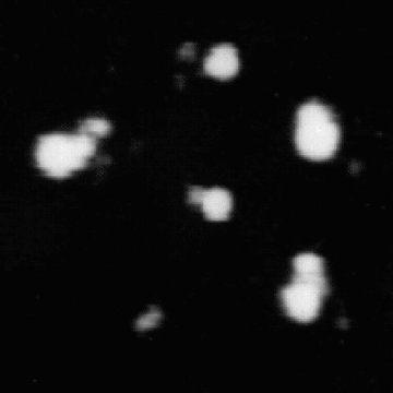
Figure 2: Experimental FIM image of an unannealed ![]() 2 nm diameter
Au cluster. Hydrogen was used as an imaging gas. (From Ref. 4).
2 nm diameter
Au cluster. Hydrogen was used as an imaging gas. (From Ref. 4).
The cluster shown in Fig. 2 was grown using a multiple
expansion cluster source (MECS). This source has the required flexibility to
grow clusters with a wide range of compositions and with a sufficient flux to
make the field-ion experiments possible. A description of this cluster source
can be found in the literature. [4, 12, 13] It is a
gas aggregation source, that is designed to run with 20 to 50 torr of
inert gas in the growth region. Both cluster growth via accretion of
single atoms and via cluster-cluster aggregation can be promoted.
A sample of the cluster aerosol produced in the MECS is expanded through
a capillary into a vacuum chamber held at ![]() Torr, resulting
in the formation of a cluster beam. As in previous studies,
[6, 7, 8, 14, 15, 16]
samples were also captured on suitable amorphous carbon grids for
further analysis by TEM. Previous TEM studies of clusters studied in
this way have confirmed the ability of the MECS to produce metal
clusters having a controlled mean size and a narrow size distribution.
[12, 13]
Torr, resulting
in the formation of a cluster beam. As in previous studies,
[6, 7, 8, 14, 15, 16]
samples were also captured on suitable amorphous carbon grids for
further analysis by TEM. Previous TEM studies of clusters studied in
this way have confirmed the ability of the MECS to produce metal
clusters having a controlled mean size and a narrow size distribution.
[12, 13]