The thin shell model [17, 18] was adopted for computer simulations in this study. This model assumes that image spots are formed by the outermost atoms on the cluster. This assumption is justified by the physical condition that tunneling of electrons from image gas atoms into the tip is related to the electric field. Since the local electric field is highest near the most protruding atoms, those atoms contribute most to the FIM image. A thin ``shell'' is defined at the surface of the cluster, and all atoms within that shell are assumed to contribute to the final image (see Fig. 3.)
To implement this technique, a cluster structure is first assumed. The
distance ![]() of each atom to the cluster's center is then computed.
This distance is then compared to the distance (
of each atom to the cluster's center is then computed.
This distance is then compared to the distance ( ![]() - t), where
- t), where
![]() is the distance from the center of the cluster to the
outermost atom and t is the thickness of a thin shell circumscribing
the cluster's perimeter. If a particular
is the distance from the center of the cluster to the
outermost atom and t is the thickness of a thin shell circumscribing
the cluster's perimeter. If a particular ![]() is greater than
(
is greater than
( ![]() - t), that atom is identified as contributing to the FIM
image and the image of that atom is projected in a way to approximate
the trajectory of ions in the field-ion microscope.
- t), that atom is identified as contributing to the FIM
image and the image of that atom is projected in a way to approximate
the trajectory of ions in the field-ion microscope.
In this study, the image is formed by a simple geometric projection of the atoms contributing to the image. The point of projection is assumed to be a distance (N+1)R from the front surface of the cluster, with R being the average radius of the cluster. With N=1 this corresponds to a stereographic projection. This standard projection is closest to typical results[19], and gives a good qualitative understanding of the expected images. In actuality, N is a function of the tip-to-screen distance as well as the angle from the apex of the tip or cluster.[19] A more accurate prediction, required for quantitative analysis of image features, would require a more intimate knowledge of the electric field surrounding the tip and supported cluster.
Each individual image spot is represented by a Gaussian intensity pattern centered about each contributing atom's projected position. The width of this Gaussian can be adjusted by the user to better match experimental images. An image spot in FIM represents a region about 0.2 - 0.3 nm in diameter, and the simulations should reflect this.
For the field-ion simulations discussed here, ideal icosahedral and truncated octahedral structures were assumed for the cluster. Crystal lattices were defined for both structures, the icosahedral cluster containing 309 atoms and the truncated octahedral cluster containing 201 atoms. These configurations correspond to clusters which are approximately 2 nm in diameter, which is about the same size as the cluster imaged in Fig. 2.

Figure 3: Schematic of the thin shell model for simulating field ion micrographs of
supported nanometer-size clusters.
The icosahedral cluster used to simulate the field-ion images was initially oriented as shown in Fig. 4 with the Z axis directed towards the screen. The truncated octahedral cluster was originally oriented as shown in Fig. 5.
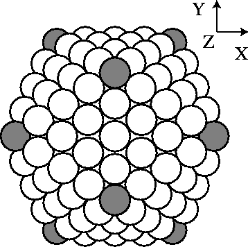
Figure 4: A 309 atom icosahedral cluster viewed with the Z axis directed at the screen. The
shaded atoms represent atoms which contribute to the field-ion
image when the shell thickness t = 0.75Å
(see Fig. 6(a).)
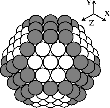
Figure 5: A 201 atom truncated-octahedral cluster viewed along [111]. The shaded
atoms
represent atoms which contribute to the field-ion image (see Fig.
7).