 |
|
|
|
|||||
| The
Group l Current
Research l Experimental
Techniques l Equipment Ejournal Links l Image Gallery |
|
||||
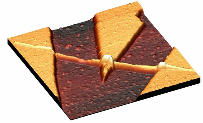
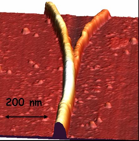
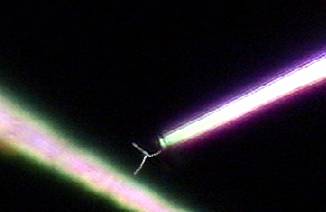
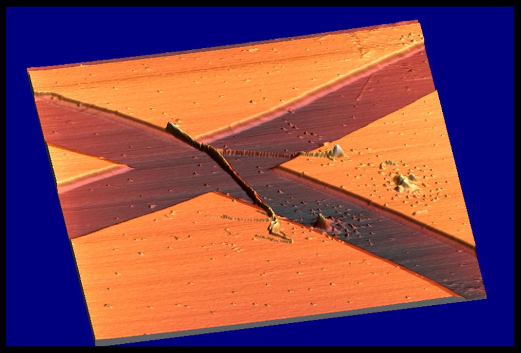
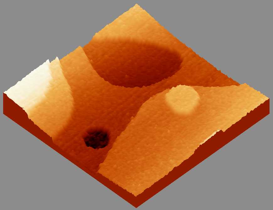
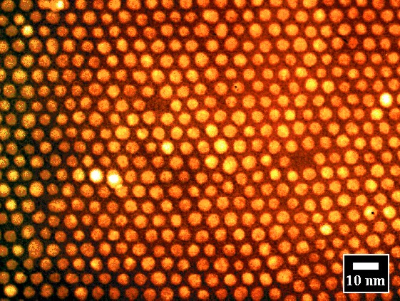
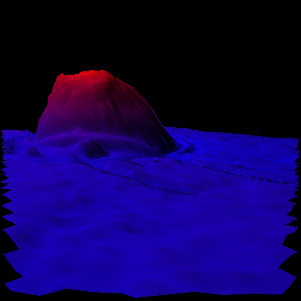
| Personnel | Scanning Probe Images |
|
 |
|
|
|||||||||||||||||||||||||||||||||||||||||||||||||||||||||||||||
Click Here
to go Back |
|||||||||||||||||||||||||||||||||||||||||||||||||||||||||||||||
|