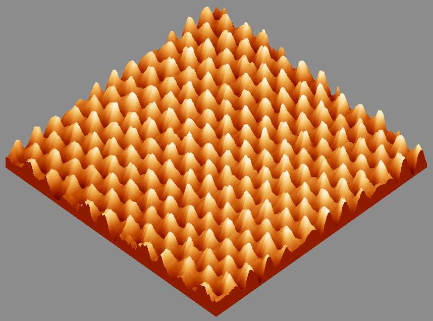
Atomic STM image of graphite (HOPG)
click to view high-resolution image
- 4 x 4 nm STM image of highly oriented pyrolytic graphite (HOPG), acquired with a tunneling current of 1.2 nA, a sample bias of -100 mV, and a scan rate of 41 Hz in the constant height mode.
- White object is carbon atom and spacing between two atoms is 2.5 angstrom.
- In this image, every other carbon atom appears from the real honey comb structure of HOPG. The resulting image is a hexagonal closed-packed pattern due to a particular symmetry of the wave functions at the Fermi surface near the K points in the surface Brillouin zone.

takhee@physics.purdue.edu
Last modified: Wed Jul 14 15:15:24 EST 1999