
David Nolte Research Group: Laser Interferometry and Holography
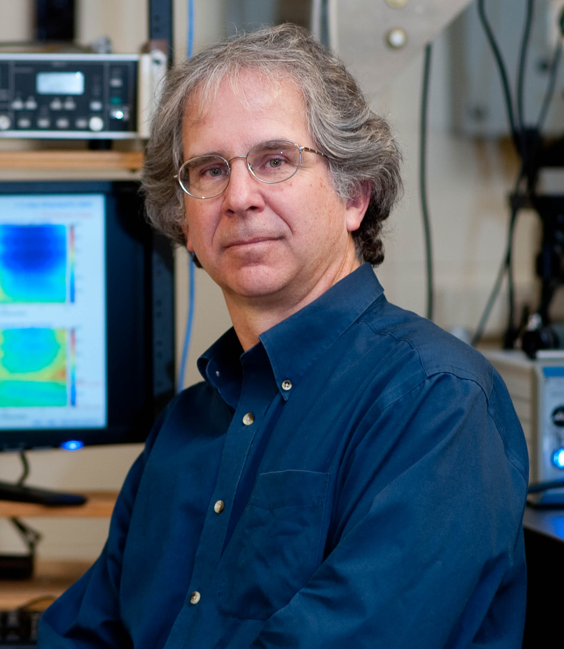
Edward M. Purcell Distinguished Professor of Physics
Professor Nolte is the Edward M. Purcell Distinguished Professor of Physics. Edward Mills Purcell (1912 - 1997) received his undergraduate BS from Purdue University in 1933 where he was encouraged by Prof. Lark-Horowitz to do undergraduate research in physics under Profs. Walerstein and Yearian. Purcell was awarded the 1952 Nobel Prize in Physics for his discovery of nuclear magnetic resonance, the physical basis of MRI imaging. In 1977 he published a landmark paper "Life at Low Reynolds Number" that applied principles of hydrodynamics to biological systems. He was awarded, with Howard Berg, the Max Delbrück Prize for biophysics of the American Physical Society in 1984. He made a lasting mark on physics education with his relativistic approach to teaching electricity and magnetism in his 1965 Berkeley Physics Course textbook.
Purcell, E. M. (1977). "Life at low Reynolds number". American Journal of Physics 45: 3–11.
Animated Dynamics (AniDyn) Inc.
Prof. Nolte is a technical founder, with John Turek of Basic Medical Sciences, of Animated Dynamics (AniDyn) Inc. that is developing Biodynamic Imaging (BDI) for early-stage drug discovery and biomedical applications that include chemotherapy selection for cancer patients and embryo selection for IVF.
Quadraspec, Inc.
Prof. Nolte is a technical founder of Quadraspec, Inc., now Perfinity Biosciences, located at the Purdue Research Park. Quadraspec developed the BioCD, a high-speed spinning biosensor disc, sold through Antech Diagnostics, capturing a 5% annual share of the US canine heartworm testing market between the years 2009 to 2021, with over 30 million dogs tested.

In the News
Dec. 11, 2025
National Academy of Inventors
Prof. David D. Nolte is elected a Fellow of the National Academy of Inventors.
Jan. 19, 2024
Comparative Oncology
First cross-species (canine and human) and cross-disease (lymphoma and esophageal caner) demonstration of biodynamic imaging. Published in Scientific Reports. Sci. Rep.
Aug 6, 2023
New Book: Interference
Interference: The History of Optical Interferometry and the Scientists Who Tamed Light published this month by Oxford University Press. Amazon Page
Dec 15, 2019
New Book: 2nd Edition: Modern Dynamics
The second edition of Modern Dynamics: Chaos, Networks, Space and Time published by Oxford University Press
Oct 15, 2018
New Book: Galileo Unbound
Galileo Unbound: A Path Across Life, the Universe and Everything published this month by Oxford University Press.
May 17, 2017
Lions Club Cancer Research Award
Prof. Nolte has been named the recipient of the 2017 Lion's Club Award for Outstanding Cancer Research at Purdue University.
Feb. 7, 2017
Outstanding Faculty Commercialization Award
Prof. Nolte was awarded Purdue University's 2017 Outstanding Faculty Commercialization Award for his entrepreneurial activities launching two biotech startups: Quadraspec and Animated Dynamics.
Sept 1, 2015
NSF Phase-II SBIR Awarded
Prof. Nolte's biotechnology start-up company, Animated Dynamics Inc., has been awarded a two-year Phase-II SBIR (Small Business Innovative Research) award from NSF to develop a Biodynamic Microscope product.
May 1, 2015
Mira Award: Indiana Technology Innovation of the Year
Animated Dynamics Inc. receives the Mira Award for Indiana Technology Innovation of the Year.
June 1, 2013
NIH R01 Grant Awarded
Purdue University, through Prof. Nolte laboratory, has been awarded an NIH R01 four-year award to study the use of Biodynamic Imaging in the selection of cancer chemotherapy in ovarian cancer. Initial studies will focus on animal models and will transition to a pilot clinical trial in human patients in 2016.
May 1, 2013
Purdue Discovery Park Fellow
Prof. David Nolte has been chosen to be a Discovery Park Fellow with joint affiliation to Bindley Bioscience Center and the Oncological Sciences Center. The project is to pursue BioDynamic Imaging across a wide range of disciplines as diverse as cancer therapy, drug discovery and development, reproductive science, developmental biology, liver and kidney disease, biomechanics and microrheology, tissue engineering, cell cycle signaling, and plant science and agriculture.
February 19, 2013
• Press Release: Animated Dynamics LLC
Animated Dynamics won first place in the 26th Burton D. Morgan Business Plan Competition. Biodynamic Imaging is a new technique that images the activity of living tissue to assesses its state of health. Applications include discovery of new anticancer drugs, selection of therapy for personalized cancer care and viability assessment for in vitro fertilization. Biodynamic Imaging measures intracellular motion through laser light scattering. Award image
November 30, 2012
• Press Release: Fellow of AAAS
David Nolte has been elected a Fellow of the American Association for the Advancement of Science (AAAS) Section on Physics
January 30, 2011
• Press Release: Tissue Dynamics Spectroscopy
• Society of Lab Automation and Screening Innovation Award Finalist
• NSF Research Nugget
Tissue dynamics Spectroscopy (TDS) uses biointerferometry to provide unique spectrogram fingerprints for the response of living tissue to applied drugs.
April, 2010
The Tangled Tale of Phase Space
Physics Today feature article traces the historical development of the concept of phase space. Physics Today Phase Space Article (PDF)
March 6, 2007
Holographic images use shimmer to show cellular response to anticancer drug
The response of tumors to anticancer drugs has been observed in real-time 3-D images using technology developed at Purdue University. (SPIE Newsroom)
October 17, 2005
David D. Nolte was the winner of the 2005 Herbert Newby McCoy Award
The Herbert Newby McCoy Award is Purdue University's highest award for outstanding contributions to science.
| McCoy Distinguished Lecture (PDF)
May 18, 2004
BioCDs could hit No. 1 on doctors' charts
While-you-wait medical tests that screen patients for thousands of disease markers could be possible with compact-disk technology patented by Purdue University scientists. The future of diagnostic blood tests may lie in your computer's CD drive -- if a Purdue scientist can carry out his vision.
May 7, 2002
Lasers light way to 3-D imaging in Purdue lab
Purdue University scientists developing a new imaging technology have created the world's first "visual fly-throughs" of a living tumor.
The Coherent Optics and Laser Interferometry Group, directed by Professor David D. Nolte uses laser interferometry to study the interaction of light with living and active matter with a special focus on the physics of partial coherence combined with the propagation of light through biological tissues. The group pioneered photorefractive quantum wells (PRQW) in the 1990's, the biological compact disk (BioCD) in the 2000's, and dynamic-contrast optical coherence tomography (DC-OCT) from 2004 to the present time. Biodynamic imaging (BDI) is based on digital holography and Doppler light scattering from intracellular motions in a physics-based approach to cancer science.
Review Articles on these topics include:
D. D. Nolte, "Semi-insulating semiconductor heterostructures: Optoelectronic properties and applications," Journal of Applied Physics 85, 6259-6289 (1999).
D. D. Nolte, "Invited Review Article: Review of centrifugal microfluidic and bio-optical disks," Review of Scientific Instruments 80 (2009).
D. D. Nolte, "Coherent light scattering from cellular dynamics in living tissues," Reports on Progress in Physics 87 (2024).
Research Positions
Research positions are open for graduate research assistantships (RAs) and for undergraduates pursuing a senior thesis. Experimental research focuses on the interaction of light and lasers with living biological systems. Computation research focuses on the application of Twin and Triplet artificial neural networks for highly variable feature classification. Theoretical research focuses on photon Monte-Carlo approaches to coherent light propagation in random dynamic media.
Holographic Biodynamic Imaging
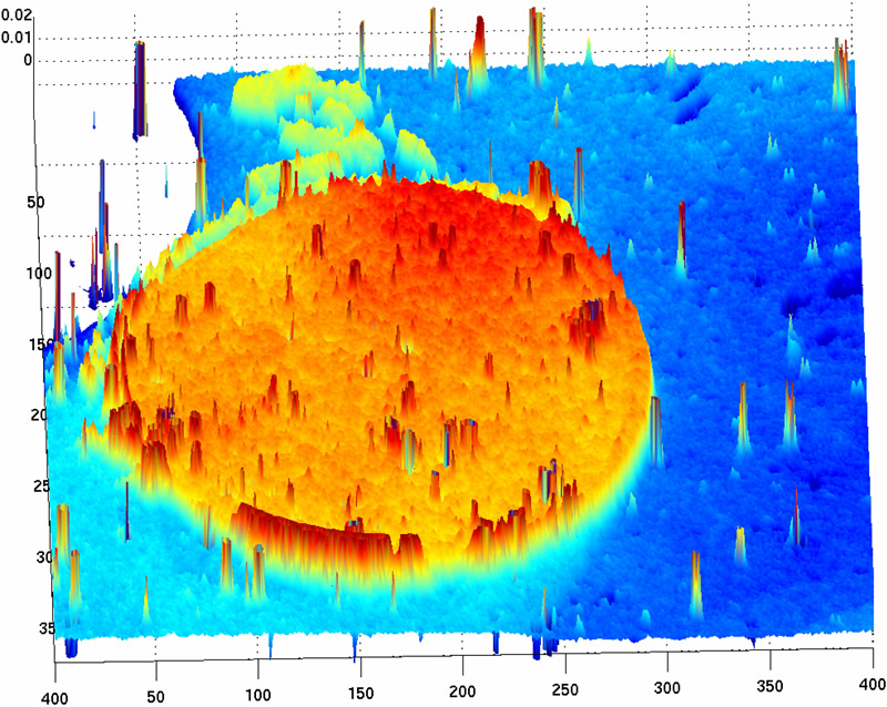
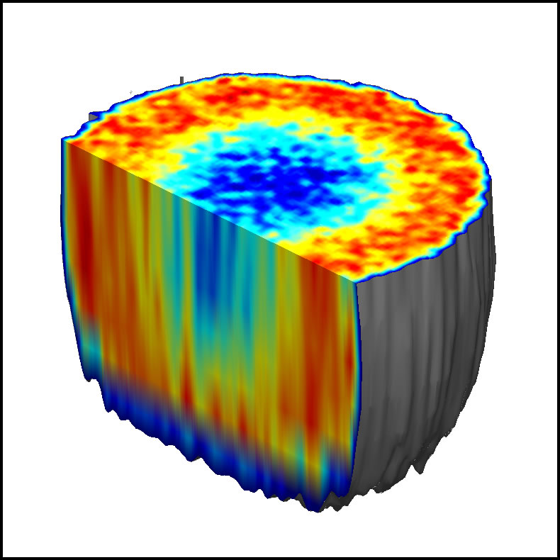 Volumetric imaging of cellular motion in a tumor spheroid. Full size
Laser fields propagating through scattering tissue retain an exponentially decaying coherence that can write holographic gratings on a holographic medium. The first volumetric holograms of living tissue were made by our group at Purdue University in 2002 using ultra-sensitive dynamic holographic film called photorefractive quantum well (PRQW) devices. Digital holography, using CCD cameras, provides similar advantages for holographic optical coherence imaging (OCI). In biomedical imaging applications, intracellular motility, recorded as shimmering holograms, acts as a novel imaging contrast agent. Recent interest is on the action of anti-mitotic cancer drugs on tissue. | Cellular motility as a novel contrast agent in digital holography of tissue
The recent development of Tissue Dynamics Spectroscopy (TDS) opens up new opportunities to study the functional influence of new drug compounds on living tissue. Tissue Dynamics Spectroscopy performs label-free non-invasive measurements of the intracellular dynamics inside living tissue and evaluates how these dynamics are altered by applied drugs. The need that TDS addresses is its ability to bridge the gap that has persisted in the drug-discovery pipeline between high-throughput 2D monolayer cultures and preclinical animal models (where drug failures are much more expensive). Three-dimensional tissue-based drug screens (for therapeutic efficacy and specificity) are needed to bridge this gap because 3D tissues are more representative of cellular and tissue responses to applied drugs than those in 2D monolayer culture. Currently there are no volumetric imaging modalities that capture drug response without labels deep in tissue far from surface effects. The response of tissue to an applied drug is represented as a drug-response spectrogram. Every different type of drug has a different spectrogram fingerprint. Different spectrogram features are related to different mechanisms of action of the drug, for instance monitoring cell metabolism or cytoskeletal integrity, or apoptotic response.
The stage in early drug discovery that comes after high-throughput screening is called “hit confirmation and expansion”. At this stage in the drug development pipeline, screens are needed to test “hits” for therapeutic efficacy (does it modify a selected target?) and specificity (does it only affect a selected target?). Target response is either molecular (pathway) or physiological (phenotypic profile). Tissue dynamics spectroscopy enters the marketplace as a new screening technology for early drug discovery. The field of use of TDS is hit confirmation and expansion. The target is high-content phenotypic profiling of physiological response to drug candidates.
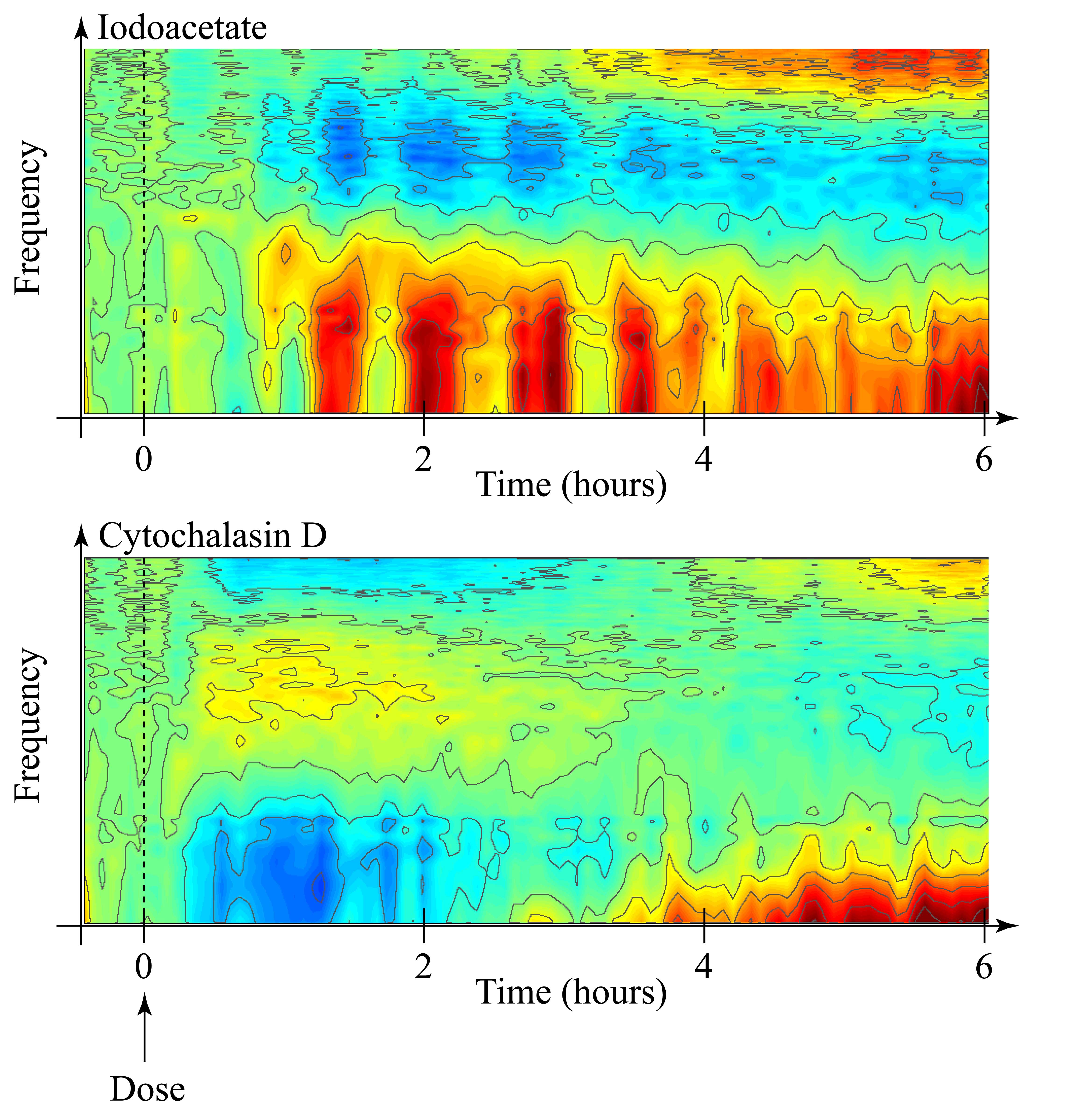
Fig. Drug Response Spectrograms for phenotypic profiling of Iodoacetate and Cytochalasin D
The BioCD
The BioCD performs molecular diagnostics in the form of a compact disc. The BioCD, invented in the Adaptive Optics and Biophotonics group at Purdue University, combines the simplicity of spinning-disc interferometry (SDI) with the power of antibodies to detect disease. A conventional compact disc has 5 billion diffraction-limited pits encoding digital information. The motivation behind the BioCD is to turn a disc into 5 billion micro-test-tubes to test for every type of blood protein in a few drops of blood. The science of molecular interferometry combines the physics of laser coherence, quantum optics, light-matter interactions, and surface science, with molecular biology and biomedical diagnostics. | Tutorial on Molecular Interferometry |
Microfluidics
Following the example of integrated electronic circuits, the "lab-on-a-chip" uses micro-fabrication to create microfluidic systems that transport liquid samples through reaction and analysis chambers for biochemical assays. We are studying the fundamental physics of fluids in micron and nanometer-scale systems. The unstable fingering of invading phases in 2D random systems, and thermodynamic properties of liquids in contact with other liquids or solids, leads to strong hysteretic relationships between saturation and capillary pressure that has eluded clear theoretical explanation. Moving into three dimensions is being pursued by building porous microsystems using two-photon polymerization (2PP) laser micromachining.
New General Interest Book: Interference and the Scientists who Tamed Light (Oxford University Press, 2023)
Available at Amazon: Amazon Author Page
Avaliable at Oxford: Link
Avaliable at Barnes and Nobles: Link
History of Physics Blog Site
Link to WordPress Blog site: Physics Blog
Selected Works
Selected publications can be found at The Berkeley Press website: Berkeley Press
David Nolte Amazon.com Author Page
Books available on Amazon.com: Amazon Author Page
Popular Science Book: Galileo Unbound: The History of the Physics of Motion (Oxford University Press, 2018)
Galileo Unbound
By David D. Nolte (Oxford University Press, 2018)
Table of Contents:
Chapter 1: Flight of the Swallows - Introduction to motion and trajectories
Chapter 2: A New Scientist - Galileos Biography
Chapter 3: Galileos Trajectory - Study of the science of motion - Publication of Two New Sciences
Chapter 4: On the Shoulders of Giants - Newtons Principia - The Principle of Least Action: Maupertuis, Euler, and Voltaire - Lagrange and his new dynamics
Chapter 5: Geometry on my Mind - Differential geometry of Gauss and Riemann - Vector spaces from Grassmann to Hilbert - Fractals: Cantor, Weierstrass, Hausdorff
Chapter 6: The Tangled Tale of Phase Space - Liouville and Jacobi - Entropy and Chaos: Clausius, Boltzmann and Poincare - Phase Space: Gibbs and Ehrenfest
Chapter 7: The Lens of Gravity - Einstein and the warping of light - Black Holes: Schwarzschilds radius - Oppenheimer versus Wheeler - The Golden Age of General Relativity
Chapter 8: On the Quantum Footpath - Heisenbergs matrix mechanics - Schrödingers wave mechanics - Bohrs complementarity - Einstein and entanglement - Feynman and the path-integral formulation of quantum
Chapter 9: From Butterflies to Hurricanes - KAM theory of stability of the solar system - Steven Smales horseshoe - Lorenz butterfly: strange attractor - Feigenbaum and chaos
Chapter 10: Darwin in the Clockworks - Charles Darwin and the origin of species - Fibonnaccis bees - Economic dynamics - Mendel and the landscapes of life - Evolutionary dynamics - Linus Paulings molecular clock and Dawkins meme
Chapter 11: The Measure of Life - Huygens, von Helmholtz and Rayleigh oscillators - Neurodynamics - Euler and the Seven Bridges of Königsberg - Network theory: Strogatz and Barabasi
Textbook on Modern Dynamics (Oxford University Press, 2nd Edition 2019)
Introduction to Modern Dynamics: Chaos, Networks, Space and Time
By David D. Nolte (Oxford University Press, 2019)
A Jr/Sr Mechanics/Dynamics textbook from Oxford University Press, updating how we teach undergraduate physics majors with increased relevance for careers in changing times. Topics: Trajectories, Geodesics, Hamiltonian Physics, Chaos, Evolutionary Dynamics, Synchronization, Dynamic Networks, Neural Networks, Economic Dynamics, Differential Geometry, Special Relativity, General Relativity
A Companion Volume to Intro to Modern Dynamics can be found at www.works.bepress/ddnolte
Textbook on BioInterferometry (Springer, Fall 2011)
 Optical Interferometry for Biology and Medicine, by David D. Nolte (Springer, 2011)
Topics: Interferometers, Speckle, Holography, Optical Coherence Tomography, Biosensors, BioCD, Cellular Dynamics, Dynamic Light Scattering, Interference Microscopy, Nanoparticles, Motility Contrast Imaging, Tissue Dynamics Spectroscopy. Table of Contents (PDF)
Link to Amazon.com
General-Interest Book on Optical Technologies at the turn of the (new) Century (Free Press, Fall 2001)
 Mind at Light Speed: A New Kind of Intellegence, by David D. Nolte (Free Press, 2001)
Genre: Science nonfiction. Topics: The Glass Bead Game, Machines of Light, Visual Communication, Telecom, Quantum Information and Quantum Computing.
Link to AbeBooks.com