The cytochrome b6f structure simulations
(S. Savikhin’s group)
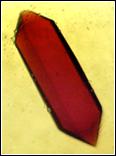
We have recently shown that the lifetime of the Chl a molecule in the cytochrome b6f complex becomes up to 5-6 times longer when protein is in a compact crystal structure like one shown below.
|
|
Single crystal of the cytochrome b6f has a size of ~20 m x 80 m and performing femtosecond pump-probe experiment on such a sample is a challenging task |
|
|
Excited state lifetime of the Chl a embedded deep within the protein become dramatically longer when in crystal form, suggesting that the local environment of Chl a is noticeably perturbed in the crystal. Since interpretation of experimental data is usually based on structure obtained for crystal forms of proteins, it is essential to know what kind of perturbation can be caused by crystal packing. |

Below we show arrangement of the cytochrome b6f complexes within each of the crystals studied:

|
|
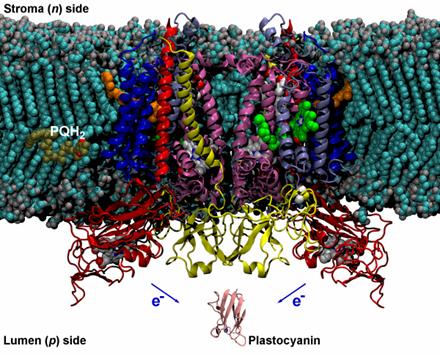
Since the structure of the cytochrome b6f in its native environment – photosynthetic membrane – cannot be determined independently, we use molecular dynamic simulations run on supercomputer in order to predict possible deviation of the native complex structure from that in crystal. The crystal structure of the complex is placed into artificially modeled lipid membrane and surrounded by water. The resulting system consists of 320,171 atoms and its structure is the optimized for minimum energy on a supercomputer. |
|
|
This is the minimized structure of the cytochrome b6f complex obtained on RCAC supercomputer using NAMD 2.5 program and CHARMM 27 force field. Download the movie showing the first 100 ps of simulation of the structure around chlorophyll a in motion |